2024年7月11日木曜日
Comparison of #Left_Ventricular #End-Diastolic_Volume Approximated from #Mean_Blood_Pressure and #Stroke_Volume and #End-Diastolic_Volume Calculated from Left Ventricular-Aortic Coupling
Comparison of Left Ventricular End-Diastolic Volume Approximated from Mean Blood Pressure and Stroke Volume and End-Diastolic Volume Calculated from Left Ventricular-Aortic Coupling
by Takahiro Shiraishi,
Department of Anesthesiology and Reanimatology, University of Fukui Hospital, Fukui 910-1193, Japan
J. Clin. Med. 2024, Accepted: 27 May 2024 / Published: 29 May 2024
Abstract
Objectives: The purpose of this study was to compare left ventricular end-diastolic volume (EDV), derived from left ventricular arterial coupling (Ees/Ea), and mean arterial blood pressure. Both of these methods of measuring EDV require some invasive procedure. However, the method of measuring EDV approximate is less invasive than the EDV coupling measuring method. This is because EDV approximate only requires arterial pressure waveform as an invasive procedure. Methods: This study included 14 patients with normal cardiac function who underwent general anesthesia. The point when blood pressure stabilized after the induction of anesthesia was taken as a baseline according to the study protocol. At the point when systolic arterial blood pressure fell 10% or more from the baseline blood pressure, 300 mL of colloid solution was administered over 15 min. EDV approximate and EDV coupling were calculated for each of the 14 patients at three points during the course of anesthetic. Each value was obtained by calculating a 5 min average. The timing of these three points was 5 min before, 5 min during, and 5 min after infusion loading. Results: The total number of comparable points was 42; 3 points were taken from each of the 14 participants. Both EDV approximate and EDV coupling increased through the infusion load testing. Scatter plots were prepared, and regression lines were calculated from the obtained values. A high correlation was shown between EDV approximate and EDV coupling (R2 = 0.96, p < 0.05). Conclusions: In patients with good cardiac function, EDV approximate can be substituted for EDV coupling, suggesting the possibility that EDV can be continuously and less invasively calculated under the situation of general anesthesia.
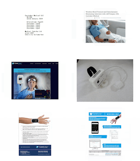
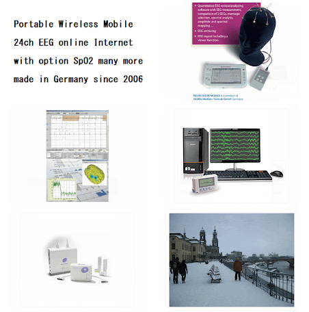
#VitalStream #Labtech_Holter #Pedcath8 #Mennen_Medical #Shimmer_sensing #Heart_Vest_gtec #wavelet_algoithm #Nevrokrd #Arteriograph #Cardionics_Stethoscope
#VitalStream_caretaker #Cardiac_Output #Artificial_Stiffness #Blood_Flow #Stroke_Volume #Care_during_after_Cardiovascular_Surgery #Labtech_Holter #Arteriograph_ArtificialStiffness #Pedcath8_MennenMedical #Cardionics_Stethoscope_SingalOutput #Nevrokard_AutonomicNerves #wavelet_algorithm


